
A Breakthrough in Intraocular Pressure Measurement: bIOP(v2)
Authors: Eliasy, A., Lopes, B.T., Wang, J., Abass, A., Vinciguerra, R., Vinciguerra, P., Bao, F.J., and Elsheikh, A.
Journal: Current Eye Research
Publication Date: Jan 2023
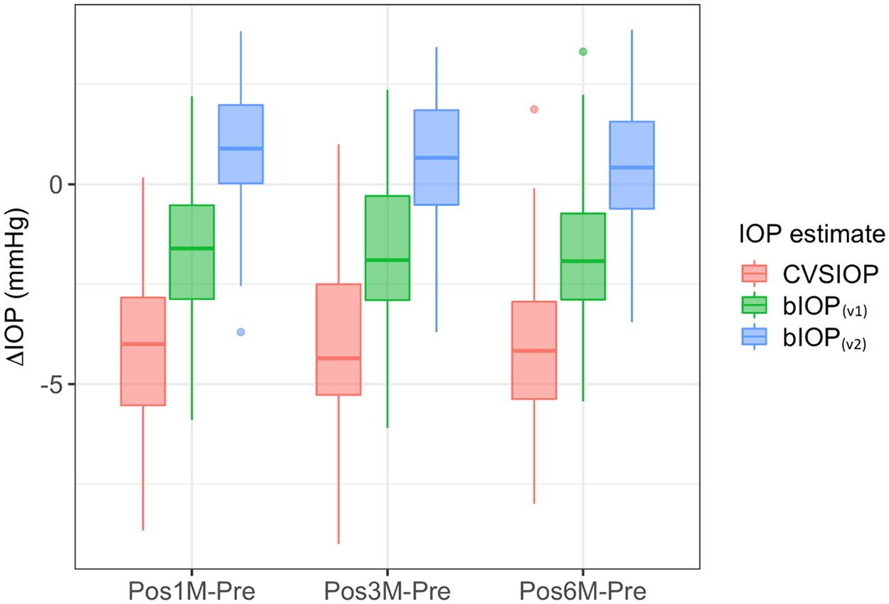
Box plot of differences between postoperative and preoperative IOP measurements in the LASIK group (box: interquartile range, bar: median).
Summary:
Intraocular pressure (IOP) plays a significant role in diagnosing and managing glaucoma, a leading cause of irreversible blindness. Existing IOP measurement methods, however, are influenced by corneal stiffness, which can lead to inaccuracies in readings. Our research team has developed an updated Biomechanically Corrected Intraocular Pressure (bIOP) measurement method, known as bIOP (v2), which aims to reduce the dependence on corneal biomechanics for more accurate IOP readings.
In the past, attempts to create more accurate IOP estimations have been made, with varying degrees of success. Our earlier study introduced the bIOP(v1) algorithm, which showed reduced influence of corneal stiffness on IOP measurements, but some correlation with stiffness parameters remained. To further improve the algorithm, we developed bIOP (v2) using improved and more representative numerical modeling, followed by clinical validation.
Our study showed that bIOP (v2) had a minimal reduction in IOP measurements post-refractive surgeries like Small Incision Lenticule Extraction (SMILE) and Laser-Assisted In Situ Keratomileusis (LASIK). This suggests that the new bIOP (v2) is less influenced by the biomechanical changes caused by these procedures, resulting in more accurate IOP measurements.
Although our study has some limitations, such as the lack of comparison with other established tonometry methods, the results are promising. The bIOP (v2) offers a more accurate and less biomechanics-dependent IOP measurement, which could potentially improve glaucoma diagnosis and management in the future.